the circulatory system
Heart Disection
External Examination of the heart
Describe the appearance of the heart. What does it look like? How does it feel? Are there any features you can describe?
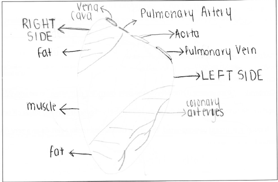
The heart was a pink- red colour and quite tough and muscular in texture. Some parts of the heart were really white because they were covered in thick amounts of fat. The coronary arteries and aorta were visible on the surface of the heart and were a dark blue-purple colour. Collectively they appeared spidery, long and thin. The visible arteries and veins in the heart were very tubular and large.
Provide a sketch of the front exterior of the heart. Label all key parts in the space below;
Find the blood vessels on the surface of the heart muscle. These are the coronary arteries. They carry nutrients and oxygen to the heart muscle.
Describe what this artery looks like.
The coronary arteries were long, thin, spidery and very dark in colour. They branched out into littler arteries around the surface of the heart starting at the aorta.
What do you think would happen if this artery was blocked by a clot?
If the coronary arteries were blocked then the blood flow to the heart would be reduced. Because red blood cells are rich in oxygen, the heart would be deprived of the oxygen it requires.
How do you know which is the left and right side of the heart?
The most accurate way to distinguish between the left and right side of the heart is to feel which ventricle has the thickest muscle wall- the thickest being the left side. Another way is to look at the atria as the left side is the biggest chamber in the heart. Furthermore the coronary arteries were only visible on the left side of this heart creating another distinguishable feature.
Have a feel of the thickness of the heart muscle at the top and bottom of the heart. Describe the following features;
The thickness of the muscles at the top of the heart
The thickness of the muscles at the top of the heart is minimal compared to that of the lower parts. It is very soft and elastic.
The thickness of the muscles at the bottom of the heart.
The muscles at the bottom of the heart are very thick and dense- especially on the left side. The bottom of the heart is very muscular and tough.
The amount of fat surrounding the heart.
In parts of the heart there was thick layers of fat condensed together. The fat was white in colour, very solid, dense and thick and when broken it crumbled.
Any major vessels entering and exiting the heart.
The major vessels of the heart which include the aorta, pulmonary artery, pulmonary vein and the vena cava were all varied in the thickness of their muscular walls. The also slightly differed in size. All of these blood vessels were long and spidery as a result of them branching out into smaller vessels like the coronary arteries.
Find the pulmonary artery that leaves the right ventricle and the pulmonary vein which enters the left atrium and complete this sentence;
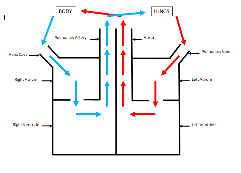
De-oxygenated blood leaves the right ventricle in an artery and travels to the lungs. Here, the blood collects oxygen, so it is now oxygenated. The blood travels back to the heart via a vein.
Find the aorta that carries the blood away from the left ventricle of the heart.
Describe the thickness of this vessel. Why do you think it needs to be so thick?
The aorta was a fairly thick tubular muscle. Its walls were very tough so that could be able to withstand the pressure of each heart beat pumping the blood through the heart and out to the rest of the bodies organs.
Where is it taking blood to?
The blood is being taken to the brain and the rest of the body.
Find the vena cava. This is the vein that returns blood from the body.
Compare the thickness of the vena cava to the aorta. Why do you think it is different?
The aorta muscle wall was much thicker than the vena cava. This is because the pressure of the heart beat from the artery is lost in the capillaries so by the time the blood reaches the veins, there is virtually a non-existent pressure fill in the vein.
What part of the heart does the vena cava go back in to?
The vena cava pumps deoxygenated blood into the right atrium, then the right ventricle and then into the pulmonary artery which takes the blood back to lungs to retain oxygen.
Remember what you observed when you observed the water flowing through the heart. The water went into the vena cava and into the heart. Which blood vessel did the water come out of the heart from?
When water entered the vena cava it came out of the pulmonary artery.
When the water was flowing into the pulmonary vein, which vessel did it come out of?
After the water entered the pulmonary vein it came out of the aorta.
Describe what this artery looks like.
The coronary arteries were long, thin, spidery and very dark in colour. They branched out into littler arteries around the surface of the heart starting at the aorta.
What do you think would happen if this artery was blocked by a clot?
If the coronary arteries were blocked then the blood flow to the heart would be reduced. Because red blood cells are rich in oxygen, the heart would be deprived of the oxygen it requires.
How do you know which is the left and right side of the heart?
The most accurate way to distinguish between the left and right side of the heart is to feel which ventricle has the thickest muscle wall- the thickest being the left side. Another way is to look at the atria as the left side is the biggest chamber in the heart. Furthermore the coronary arteries were only visible on the left side of this heart creating another distinguishable feature.
Have a feel of the thickness of the heart muscle at the top and bottom of the heart. Describe the following features;
The thickness of the muscles at the top of the heart
The thickness of the muscles at the top of the heart is minimal compared to that of the lower parts. It is very soft and elastic.
The thickness of the muscles at the bottom of the heart.
The muscles at the bottom of the heart are very thick and dense- especially on the left side. The bottom of the heart is very muscular and tough.
The amount of fat surrounding the heart.
In parts of the heart there was thick layers of fat condensed together. The fat was white in colour, very solid, dense and thick and when broken it crumbled.
Any major vessels entering and exiting the heart.
The major vessels of the heart which include the aorta, pulmonary artery, pulmonary vein and the vena cava were all varied in the thickness of their muscular walls. The also slightly differed in size. All of these blood vessels were long and spidery as a result of them branching out into smaller vessels like the coronary arteries.
Find the pulmonary artery that leaves the right ventricle and the pulmonary vein which enters the left atrium and complete this sentence;
De-oxygenated blood leaves the right ventricle in an artery and travels to the lungs. Here, the blood collects oxygen, so it is now oxygenated. The blood travels back to the heart via a vein.
Find the aorta that carries the blood away from the left ventricle of the heart.
Describe the thickness of this vessel. Why do you think it needs to be so thick?
The aorta was a fairly thick tubular muscle. Its walls were very tough so that could be able to withstand the pressure of each heart beat pumping the blood through the heart and out to the rest of the bodies organs.
Where is it taking blood to?
The blood is being taken to the brain and the rest of the body.
Find the vena cava. This is the vein that returns blood from the body.
Compare the thickness of the vena cava to the aorta. Why do you think it is different?
The aorta muscle wall was much thicker than the vena cava. This is because the pressure of the heart beat from the artery is lost in the capillaries so by the time the blood reaches the veins, there is virtually a non-existent pressure fill in the vein.
What part of the heart does the vena cava go back in to?
The vena cava pumps deoxygenated blood into the right atrium, then the right ventricle and then into the pulmonary artery which takes the blood back to lungs to retain oxygen.
Remember what you observed when you observed the water flowing through the heart. The water went into the vena cava and into the heart. Which blood vessel did the water come out of the heart from?
When water entered the vena cava it came out of the pulmonary artery.
When the water was flowing into the pulmonary vein, which vessel did it come out of?
After the water entered the pulmonary vein it came out of the aorta.
Internal examination
Firstly cut open the LEFT VENTRICLE following the lines on the diagram above.
Describe what you see inside the left side of the heart.
The left side of the heart has very thick muscular walls around it. It is a very bright pink colour and has lots of blood around it. The left atrium was slightly larger than the ventricle and in between both chambers were long and stringy looking valves. The valves serve a purpose to stop blood flowing back into the heart the wrong way.
Observe any valves you see. What do you think their job would be?
The valves in the left side of the heart are quite thin, elastic and long. This appearance and texture coincides with its purpose which is to open and close depending on the direction of blood flow. The reason that the valves open and close is so that the blood doesn’t start flowing downwards and pooling when it should be flowing upwards.
Cut the aorta. Describe how it appears and how it feels and any other features
The aorta feels very muscular, tough and elastic. The aorta is very long and fairly wide so it can pump blood to the rest of the body.
Cut open the RIGHT VENTRICLE and observe this side of the heart. Furthermore, Cut into the atria of both the left and right chambers so you can see the muscle thickness of all four chambers.
In the table below, indicate the location and role of the following structures;

box heart diagram
Walking the heart
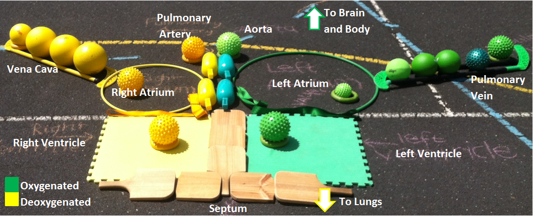
In order to develop an extended understanding of the process by which the heart pumps blood around itself and the rest of the body, we developed an abstract heart using sports equipment and simulated oxygenated blood with the colour green and deoxygenated blood with the colour yellow. The ventricles were created using foam sheets and the atria using hula hoops. The spiky and smooth surfaced balls throughout the diagram are used to demonstrate the blood flow.
Our group demonstrated the process of blood moving through the heart. We motioned how; deoxygenated blood enters the right atrium via the vena cava collected from the rest of the body (upper and lower). It then travels through to the right ventricle where it is pushed up and into the pulmonary artery which transports the blood to the lungs to gain oxygen. Once the blood is oxygenated it then travels via the pulmonary vein to the left side of the heart so it can be taken to the rest the body. Once the blood is in the left atrium it is pumped to the left ventricle then outwards into the aorta which is the largest artery in the body. The aorta does its job to push the blood to places like the brain and major bodily organs before the process repeats itself again. Within the time space of a minute the body can pump its entire blood volume around itself.
Our group demonstrated the process of blood moving through the heart. We motioned how; deoxygenated blood enters the right atrium via the vena cava collected from the rest of the body (upper and lower). It then travels through to the right ventricle where it is pushed up and into the pulmonary artery which transports the blood to the lungs to gain oxygen. Once the blood is oxygenated it then travels via the pulmonary vein to the left side of the heart so it can be taken to the rest the body. Once the blood is in the left atrium it is pumped to the left ventricle then outwards into the aorta which is the largest artery in the body. The aorta does its job to push the blood to places like the brain and major bodily organs before the process repeats itself again. Within the time space of a minute the body can pump its entire blood volume around itself.
